

What is biological neural network?
Artificial Intelligence & Machine Learning

A biological neural network is a collection of interconnected neurons that work together to process and transmit information throughout the body.
The human brain is an example of a biological neural network!
Furthermore, similar networks can be found in other animals as well.

These networks are highly complex and can perform a wide range of tasks, from simple reflexes to complex cognitive processes like decision-making and language processing.
In contrast, artificial neural networks (ANNs) are computational models inspired by the structure and function of biological neural networks.
ANNs are composed of a large number of interconnected processing units, called neurons, that are organized into layers. Each neuron receives input from other neurons and applies a non-linear function to the input. Producing an output signal sent to other neurons in the next layer. The connections between neurons become typically weighted. Allowing the network to learn from examples and adjust its behavior accordingly.
While biological and artificial neural networks share some similarities, there are also several key differences between them.

Biological neurons are highly specialized cells performing a wide range of functions. While artificial neurons are much simpler and are typically designed to perform a specific task.
Biological neural networks are highly parallel.

Meaning that many neurons can work together at the same time to process information!
In contrast, most artificial neural networks are highly sequential, meaning that the neurons process information one at a time!

Biological neural networks are highly adaptive and can learn from experience, while most artificial neural networks require a training dataset to learn from.
By studying the structure and function of biological neural networks, researchers can continue to improve the design and performance of artificial neural networks, leading to even more advanced and powerful AI systems in the future.


One of the earliest pioneers in the field was the Spanish neuroanatomist Santiago Ramón y Cajal.

Cajal used a microscope to examine the structure of the brain and nervous system in detail. He discovered that the brain became composed of discrete cells called neurons. Connected to each other by specialized junctions called synapses.
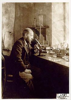
Cajal proposed the idea that information becomes transmitted through the nervous system! As a result of electrical and chemical signals traveling from one neuron to another.

Another key figure in the development of our understanding of biological neural networks was the Canadian neurophysiologist Wilder Penfield.

In the 1930s and 1940s, Penfield conducted a series of experiments in which he stimulated different areas of the brain with electrical currents while patients were awake and conscious. This allowed him to map the different regions of the brain and gain insights into how they functioned.
In the decades that followed, researchers continued to study the structure and function of biological neural networks in more detail.


In the 1950s and 1960s, for example, the British neurophysiologist Alan Hodgkin and the Australian biophysicist Andrew Huxley developed a mathematical model of the action potential, the electrical signal that travels along the axon of a neuron.

This model helped to explain how neurons transmit information over long distances!
In the 1970s and 1980s, researchers began to use computers to model biological neural networks and study their behavior. One key breakthrough during this time was the development of the backpropagation algorithm, which allowed artificial neural networks to learn from experience and adjust their behavior accordingly.
Today, research on biological neural networks continues to advance rapidly, aided by advances in technology like fMRI and optogenetics. This research is helping to shed new light on how the brain works, and is also driving the development of more advanced and sophisticated artificial neural networks.
fMRI stands for functional magnetic resonance imaging.
It is a non-invasive imaging technique that allows scientists and clinicians to study brain activity by measuring changes in blood flow in response to different tasks or stimuli.
fMRI works by using strong magnetic fields and radio waves to measure changes in the oxygen levels of blood in different parts of the brain. When a particular region of the brain becomes active, it requires more oxygen and glucose to fuel the increased metabolic demand of the neurons in that area. As a result, blood flow to that area increases, delivering more oxygenated blood and removing deoxygenated blood. These changes in blood flow can become detected by the fMRI scanner. As a result used to create a map of brain activity.
One of the key advantages of fMRI is that it allows researchers to study brain activity in real-time while subjects perform different tasks or experience different stimuli. This can provide insights into the neural mechanisms underlying complex cognitive processes like attention, memory, emotion, and decision-making.
Optogenetics is a cutting-edge technique that combines genetic engineering and optics to control specific cells in living tissues, such as neurons in the brain.
It involves the use of light-sensitive proteins called opsins, which are expressed in the target cells using genetic manipulation. These opsins can be activated or inhibited by different wavelengths of light, allowing researchers to selectively turn them on or off using fiber-optic cables or other light sources.
In the context of neuroscience, optogenetics becomes used to study the function of neural circuits. And the relationship between specific groups of neurons and behavior. Furthermore, by using optogenetics to activate or inhibit specific neurons in the brain, researchers can observe how this affects behavior and cognitive function. For example, they can study the role of specific neurons or neural circuits in processes like learning and memory, decision-making, or perception.
Optogenetics has several advantages over other methods of manipulating neurons.
Such as electrical stimulation or chemical manipulation. It is highly selective, meaning that researchers can target specific cells or circuits with a high degree of precision. It is also reversible, allowing researchers to turn cells on or off in a controlled manner over time. Additionally, because it uses light, optogenetics can be used to manipulate cells in a non-invasive way, making it ideal for use in animal models and potentially even in humans in the future.
Optogenetics has a wide range of potential applications, both in neuroscience and in other fields such as medicine and biotechnology. For example, it could be used to develop new treatments for neurological and psychiatric disorders, by allowing researchers to selectively stimulate or inhibit specific brain regions or circuits. It could also be used to engineer cells for therapeutic purposes, such as creating insulin-producing cells for diabetes treatment.
Artificial Intelligence & Machine Learning
What is biological neural network?


